Abstract
Background:
Diffuse large B-cell lymphoma (DLBCL) is a highly heterogeneous disease with variable clinical and molecular features. Studies have highlighted the significant role of γδ T cells in the survival of leukemia patients. However, the heterogeneity of γδ T cells and their impact on clinical correlation in the peripheral blood of patients with DLBCL remain unclear.
Method:
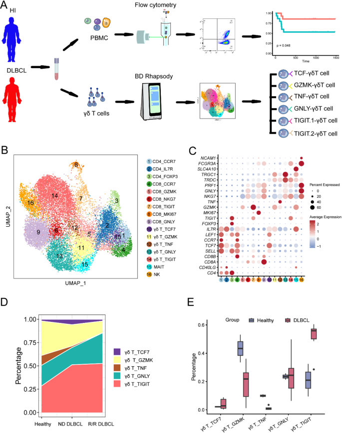
Single-cell RNA sequencing (scRNA-seq) was employed on 9 blood samples, sourced from 6 patients with diffuse large B-cell lymphoma (DLBCL) and 3 healthy individuals (HIs), to delineate clinically pertinent γδ T cell states and subsets in DLBCL patients. Flow cytometry was then employed to validate the relationship between DLBCL prognosis and γδ T cell subsets.
Result:
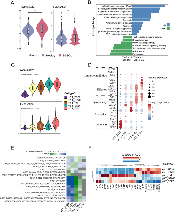
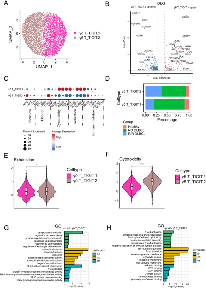
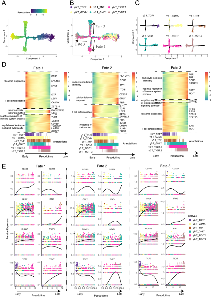
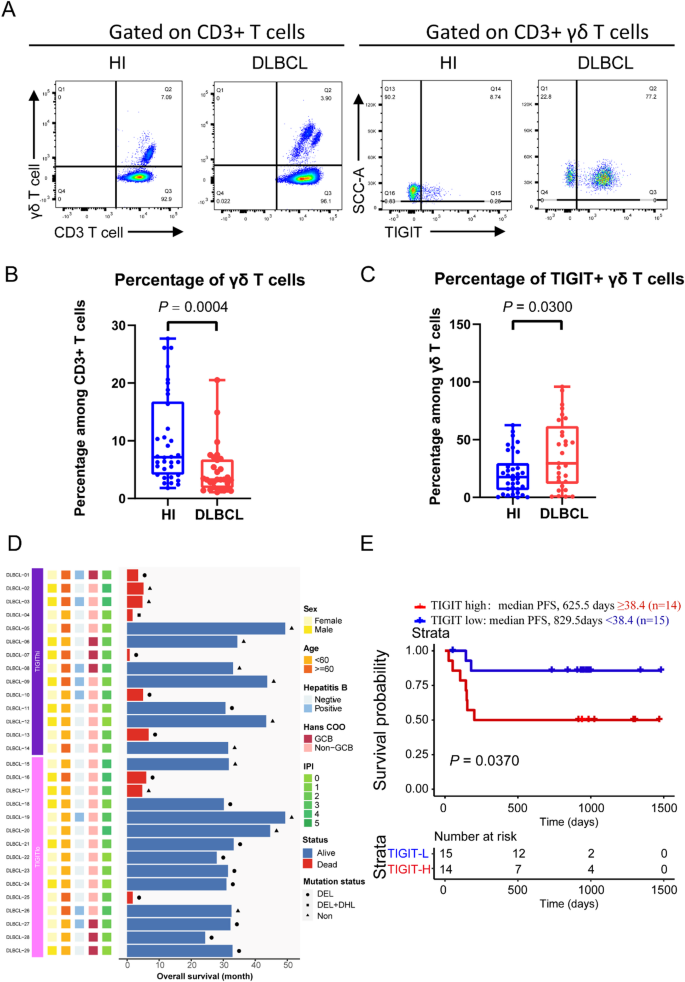
Our study integrated genetic drivers through consensus clustering, leading to the identification of 6 distinct γδ T cell subsets in DLBCL and HIs. These subsets include a naïve γδ T cell subset characterized by TCF7 and LEF1 expression, a memory γδ T cell subset sharing common genes such as GZMK, IL7R, an anti-tumor γδ T cell subset with overexpression of IFNG, TNF, and CD69, and two subsets exhibiting TIGIT overexpression indicative of an exhausted γδ T cell phenotype. Additionally, a cytotoxic γδ T cell subset marked by increased NKG7 and GZMB levels was identified. Our results revealed that while γδ T cells possess anti-tumor capacities, their functional effectiveness is diminished due to differentiation into exhausted subpopulations. Several clusters with high cytotoxicity scores also showed elevated exhaustion scores (C13-γδ-TIGIT.1, C14-γδ-TIGIT.2), suggesting the presence of a population in DLBCL samples that is simultaneously exhausted and cytotoxic. In particular, the TIGIT.2 γδ T cell subset manifests a more pronounced exhaustion score relative to TIGIT.1 γδ T cell subset, indicating differential levels of cellular exhaustion among these groups. Our analysis reveals a significant correlation between high expression of TIGIT γδ T cell subsets and poorer patient prognoses. We also discovered unique expression profiles within these subgroups: TIGIT.1 γδ T cells are marked by elevated CXCR4 expression, contrasting with the TIGIT.2 γδ T cell subgroup which exhibits increased CX3CR1 expression. Pseudotime analysis implies a potential differentiation trajectory from naïve and GZMK γδ T cells to various terminally differentiated subsets, with genes associated with stemness (e.g., TCF-1) subsequently downregulated. These findings suggest that TIGIT.2 subset may be further along in the differentiation trajectory, potentially representing a more terminally differentiated state than TIGIT.1 subset. According to our clinical validation cohort, the TIGIT+ γδ T cell subset is highly expressed in patients and correlates with poor prognosis.
Conclusion:
We identified genetic subtypes of γδ T cells with distinct genotypic and clinical characteristics in DLBCL patients. Expression levels within these subgroups emerged as potential indicators for patient outcomes and as crucial factors in shaping therapeutic strategies. These insights significantly advance our understanding of intricate relationships among cellular subgroups and their roles in influencing disease progression and patient prognosis.